

Dr. Jho's Anterolateral or Posterolateral Approach for Spinal Cord Tumors: Minimally Invasive Spinal Cord Tumor Operation
Home: Dr. Jho's Innovative Minimally Invasive Neurosurgery for Spine and Brain Disorders
Facts About These Surgeries
Discussion
Both of these approaches, the anterolateral approach which means the surgeon approaches from the front and to the side, and the posterolateral approach which indicates the surgeon is working from the back and to the side, are bone sparing procedures which focus on maintaining the structural stability of the spine. Conventional procedures often require extensive bone removal from the spine which then necessitates bone fusion and or the placement of metal implants to provide stability. As an analogy, it would be like planing, shaving, and sanding the wood on the legs of a table until it was unable to stand without additional support. Therefore, continuing with the analogy, a person undergoing one of Dr. Jho's procedures could be likened to a table requiring intricate leg work that afterwards stands as steady as it did before the work was done. The anterolateral approach spares bone by using anterior foraminotomy holes that are small and do not weaken the bone when made for tumor access. The posterolateral approach spares bone by utilizing a hemilaminectomy. Most patients are able to be discharged the day following surgery.
Definitions
foraminotomy - enlargement of an opening or passageway for a nerve
hemilaminectomy - excision of a vertebral lamina on one side
Images
Anterolateral Approach to Spinal Cord Tumors
Schematic drawings demonstrate exposure of a spinal cord tumor through a small hole of anterior microforaminotomy (anterolateral approach).


An axial view of an MRI scan shows a spinal cord tumor (left) and complete resection of the tumor is noted in the postoperative scan (right).


MR scans, sagittal view, reveal a spinal cord tumor and bony spurs at the C5-6 level preoperatively (left) and total removal of the tumor and bony spurs postoperatively (right). In this technique, bone graft fusion is not necessary and not used. Patients are hospitalized overnight. Patients do not need cervical collars postoperatively.


Intraoperative photographs taken during an anterolateral approach to a spinal cord tumor
Posterolateral Approach for Spinal Cord Tumors


Schematic drawings show a patient's positioning, a line of a skin incision (3-5cm) and a surgical approach.
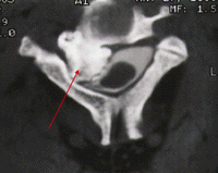

A bony tumor is demonstrated in a postmyelographic CT scan preoperatively (left, arrow) and total removal of the tumor is confirmed in a postoperative CT scan (right).


A preoperative MR scan, sagittal view, reveals a spinal cord tumor (left, arrow). A postoperative MR scan, sagittal view, confirms total removal of the tumor (right).

An intraoperative photograph demonstrates the lateral aspect of the spinal cord and the tumor (arrows).
References
Jho, HD and Ha, HG: Anterolateral approach via an anterior foraminotomy for cervical spinal cord tumor: Technical note. Minimally Invasive Neurosurgery
Jho HD: Posterolateral approach for cervical spinal cord tumor: Technical note. Journal of Neurosurgery