Dr. Jho's Anterior Microforaminotomy
for Cervical Disc Herniation: A Minimally Invasive Cervical Disc-Preserving functional
Operation - Neck Surgery
Disc-preserving outpatient surgery
Home: Dr. Jho's Innovative Minimally Invasive Neurosurgery for Spine and Brain Disorders
![]() Facts About This Surgery
Facts About This Surgery
Discussion
The focus of this newly developed surgery centers on removal of the bony spur or disc material that is pressing on the nerve root causing "electric", "numbing", or "shooting" pains, that often start in the neck and travel into one or both arms. Through an inch long incision at the front of the neck, the "source of the problem" (the disc material or bony spur) can be clearly seen through a small hole made at the side of the intervertebral disc, and is trimmed away leaving the majority of disc material and bone undisturbed. By doing so, which is a less invasive method than the conventional procedure which eventually leads to fusion of the bone, the motion segment or "neck joint" is preserved. Patients are most often able to go home the day of or the day after surgery, with noticeable relief from the nerve pain in their arms, and without neck braces or collars. Candidates for this surgery include patients who have a herniated cervical disc or bony spurs pinching the nerve root causing neck pain and radiating pain (radicular) in one or both arms not relieved by at least three weeks of conservative treatment. This does not necessarily exclude patients who have undergone previous neck surgeries.
Definitions
radicular - pertaining to pain caused by a nerve
foraminotomy - enlargement of an opening or passageway for a nerve
Images


Left: A preoperative MR scan, axial view, reveals disc herniation (arrow).
Right: A postoperative MR scan, axial view, demonstrates excellent removal of the herniated portion of the disc and bony spurs.


A preoperative axial view MR scan shows nerve root compression by disc material (arrow, left), and a postoperative MR scan displays a widened nerve root canal after disc removal (right).

An intraoperative picture shows the decompressed nerve root (long arrows) from its origin at the spinal cord (short arrows) to the exit site behind the vertebral artery.


(First Image) : A postoperative MR scan, sagittal view, depicts preservation of the remaining disc at C5-6 (arrow).
(Second Image) : A lateral x-ray done postoperatively reveals the opened neural foramen (arrows).
(Third Image) : The opened nerve passage can be viewed in this anteroposterior view x-ray (arrows).


Postoperative cervical flexion and extension x-rays demonstrate maintenance of the motion segment as well as spinal stability following foraminotomy.
References
Jho, HD: Microsurgical anterior cervical foraminotomy for radiculopathy : A new approach to cervical disc herniation. Journal of Neurosurgery 84:155-160, 1996
Jho, HD: Anterior microforaminotomy for cervical radiculopathy: A disc preservation technique. In Rengachary SS, Wilkins RJ (eds), Neurosurgical Operative Atlas, Williams & Wilkins Vol.7, 43-52, 1998
Jho, HD: Anterior cervical microforaminotomy. In Kang JD (guest ed), Fu F (ed), Current Techniques in Cervical Spine Surgery, Operative Techniques in Orthopeadics 8(1): 46-52, 1998 W.B. Saunders
Dr. Jho's Spinal Cord Decompression
via Anterior Microforaminotomy for Spondylotic Cervical Stenosis or Ossified
Posterior Longitudinal Ligament (OPLL) : Minimally Invasive Cervical Stenosis
Surgery - Neck Sugery
Home: Dr. Jho's Innovative Minimally Invasive Neurosurgery for Spine and Brain Disorders
![]() Facts About
This Surgery
Facts About
This Surgery
Discussion
Patients who have narrowed spinal cord canals in the neck region often suffer from numbness, weakness, pain in the arms, difficulty controlling urination and possibly difficulty with walking. Compared to conventional procedures which may lack in providing access to the front portion of the canal (laminectomy technique), or may require excessive bone removal and fusion (anterior discectomy or vertebrectomy technique), this new minimally invasive technique does not significantly weaken the structures of the cervical spine but still accomplishes enlargement of the canal. In simple terms, it cleans out a spinal cord canal that has bone build-up putting pressure on the spinal cord, so that the canal may then be likened to a water pipe without accumulated inner debris. It does so by the use of small holes, made with the use of the operating microscope or an endoscope, and are called foraminotomy holes. Another useful analogy that helps explain this procedure would be to picture removing the contents of a watermelon through a hole without cutting the watermelon apart. Once the surgery is done, patients can usually go home the following day without collars or braces. Recovery usually covers the time span of 4 to 6 weeks.
Definitions
foraminotomy - enlargement of an opening or passageway for a nerve
stenosis - a narrowing of a canal
spondylotic - refers to spondylosis which is a non-inflammatory disease of the spine that can include excess bone growth
Images


Left, axial view, and right, lateral view: Schematic drawings demonstrate decompression of the spinal cord through a small hole - the anterior microforaminotomy - leaving the majority of the cervical disc intact.

A schematic drawing demonstrates multilevel decompression via multiple foraminotomy holes.


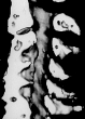
MR scans, sagittal view, demonstrate cervical stenosis at C3-4, C4-5, C5-6 preoperatively (left) and subsequent excellent decompression of the spinal cord postoperatively (right). In this technique, bone graft fusion is not necessary and not used.


A preoperative axial view MRI reveals severe spinal cord compression by a spondylotic bony spur (left). A postoperative CT scan, axial view, shows an adequately widened spinal cord canal and decompressed spinal cord (right).

A postoperative CT reconstruction depicts spinal canal decompression (left). Three foraminotomy holes (arrows) are demonstrated in this oblique reconstruction (right).


Intraoperative photographs showing spinal cord as seen through foraminotomy holes during surgical decompression.
References
Jho, HD: Decompression via microsurgical anterior foraminotomy for cervical spondylotic myelopathy. Journal of Neurosurgery 86:121-126, 1997
Jho, HD: Spinal cord decompression via microsurgical anterior foraminotomy for spondylotic cervical myelopathy. Minimally Invasive Neurosurgery 40:124-129,1997
Jho, HD: Treatment of spondylotic cervical myelopathy via anterior foraminotomy. In Camins MB (guest ed), Loftus CM, Batjer HH (eds), Cervical Spinal Stenosis, Techniques in Neurosurgery, Lippincott-Raven (in press)
For referral information or appointment for consultation contact:
Manager: Robin A. Coret, B.S.- (412)359-6110 or e-mail at
Fax: (412)359-8339
Address: JHO Institute for Minimally Invasive Neurosurgery
7th Floor, Snyder Pavilion, Allegheny General Hospital, 320 East North Avenue, Pittsburgh, PA 15212-4772
Contact Dr. Jho via email: DrJho@DrJho.com