

Dr. Jho's Cranial Nerve Surgery: Minimally Invasive Neurosurgery for Hemifacial Spasm and Trigeminal Neuralgia
Microvascular Decompression and Glycerol Rhizotomy
Home: Dr. Jho's Innovative Minimally Invasive Neurosurgery for Spine and Brain Disorders
Facts About These Surgeries
Microvascular decompression is surgical treatment for cranial nerve disorders such as trigeminal neuralgia, hemifacial spasm, vertigo, tinnitus, glossopharyngeal neuralgia and spasmodic torticollis
Glycerol rhizotomy offers a less invasive treatment for trigeminal neuralgia and is done on an outpatient basis
Discussion
Cranial nerve surgery is done through a small skull opening behind the ear and is referred to as retromastoid craniectomy. Skin incisions are usually less than two inches in length. When blood vessels cross and compress cranial nerves, various characteristic symptoms develop depending upon which cranial nerves is being compressed.. Trigeminal neuralgia develops by blood vessel compression on the trigeminal nerve, hemifacial spasm by compression on the facial nerve, intractable positional vertigo by compression on the vestibular nerve, tinnitus by compression on the cochlear nerve, glossopharyngeal neuralgia by compression on the glossopharyngeal nerve and spasmodic torticollis by pressure on the spinal accessory nerve and upper cervical nerves. Microvascular decompression which consists of placement of small synthetic sponges between the compressing blood vessels and the cranial nerve, carries a good chance of relieving those symptoms.
For frail or elderly patients, percutaneous glycerol rhizotomy is a minimally invasive alternative treatment for trigeminal neuralgia. It consists of injection through the patient's cheek of a drop or two of glycerol around the trigeminal nerve at the skull base with a spinal needle. This operation is performed under local anesthesia with sedation at the operating room and is done as outpatient surgery.
Images
An artistic depiction shows needle placement during glycerol rhizotomy (left). A flouroscopic image taken during procedure shows needle placement through the foramen ovale (right).

This intraoperative, flouroscopic image demonstrates appropriate glycerol placement (arrow) into the trigeminal cistern.

A non-contrasted axial CT scan confirms placement of glycerol in the trigeminal cistern (arrow).

For microvascular decompression, a 4-cm-long skin incision is made behind the patient's ear..


Magnified images during surgery show an arterial loop compressing the 5th cranial nerve (left, TN: trigeminal nerve), separation of the artery from the nerve (center), and placement of Teflon sponge (T) between the artery and the nerve (right).


This picture, taken through an operating microscope, demonstrates a vein (V) compressing the 8th cranial nerve (VIII) in a patient with vertigo (left). The vein is eliminated for the decompression of the 8th nerve (right, VIII).


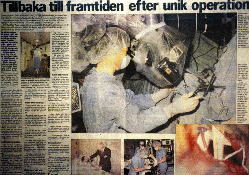
Left: This intraoperative image shows an artery compressing the 8th nerve causing intractable tinnitus (artery is located between the 8th and 7th cranial nerves). Center: 8th nerve is decompressed by placement of Teflon sponge. Right: Patient illustration as seen in a newspaper.

An intraoperative photograph displays arterial compression of the facial nerve in a patient with hemifacial spasm.


Left: An arterial loop (PICA) is progressively decompressed with Teflon placement in a patient with glossopharyngeal neuralgia. Right: Teflon placement at the completion of microvascular decompression of the ninth and tenth cranial nerves.
Reference
Jho HD, Lunsford LD: Percutaneous retrogasserian glycerol rhizotomy. In Neurosurgical perspectives on trigeminal neuralgia. Brown JA (guest ed), Neurosurgery Clinics of North America 8(1):63-74, January 1997
For referral information or appointment for consultation contact:
Manager: Robin A. Coret, B.S.- (412)359-6110 or e-mail at
Fax: (412)359-8339
Address: JHO Institute for Minimally Invasive Neurosurgery
7th Floor, Snyder Pavilion, Allegheny General Hospital, 320 East North Avenue, Pittsburgh, PA 15212-4772
Contact Dr. Jho via email: DrJho@DrJho.com